Tag Archives: physiotherapist in Dwarka

Dislocation-of-the-Patella

Physiotherapy in the Dislocation of the Patella

The patellar luxation is a disease of the locomotor system which generally occurs toward the lateral side of the knee, resulting in breakage of the medial patellofemoral ligament (LPFM) in approximately 90-100% of cases.

Although several risk factors have been identified that cause patellofemoral instability after patellar dislocation, appropriate therapy remains a controversial issue.

In this article, we will only talk about the conservative treatment of this lesion.

What is kneecap dislocation?

Label dislocation is defined as the complete loss of contact between the articular surfaces of the patellofemoral joint and in which to restore the normality of the joint a dislocation reduction by a professional with the appropriate knowledge is required, although in In certain cases this reduction occurs spontaneously through a complete extension of the knee.

Types of patellar dislocation: We found three types of patellar dislocation;

  • Congenital laxation, which occurs during birth.
  • Recurrent dislocation, which tends to recur. A dislocation is considered recurrent when it occurs two or more times.
  • Traumatic dislocation, in which there is a loss of contact between articular surfaces in an acute or traumatic way as a result of trauma or forced movement.

The elements involved in the extensor apparatus of the knee are the patella, the femoral trochlea, the quadriceps muscle, the patellar tendon, and the patellofemoral retinaculum.

In the medial retinaculum is the most important ligament in this lesion, the medial patellofemoral ligament (LPFM). This ligament consists of a transverse and an oblique portion and both portions are fused with the vast middle of the quadriceps to be inserted into the patella. This ligament is the main passive stabilizer before the dislocation of the patella during flexion from 0 to 30º.

  • The quadriceps angle (angle Q) is a measure of alignment between the femur and the tibia that is measured by drawing an imaginary line that connects the center of the label with the ESIA and a second line that is drawn following the direction of the patellar tendon, from the center of the label to the anterior tibial tuberosity Where these lines intersect, angle Q is formed.

This angle is important since its increase affects the patellofemoral stability and the path of the patella in the trochlea during knee flexion and extension.

  • The main risk factors are anatomical, and they are patellar dysplasia, the syndrome of the high patella and the increased distance between the anterior tuberosity of the tibia and the intertrochlear groove (TAT-SIT distance). However, there are other anatomical features that favor patellar dislocation such as ligament hyperlaxity, increased Q angle or atrophy or imbalance of the vast middle of the quadriceps with respect to the vast lateral and a shortened iliotibial waistband.
  • The mechanism of injury can be direct, when it is caused by trauma to the medial part of the patella or indirectly when it is caused by a knee flexion movement along with with a knee valgus and internal rotation adduction of the femur, while the tibia abducts and rotates externally.

The indirect mechanism represents 93% of the cases of the dislocated patella.

  • For the evaluation and diagnosis of the lesion, in addition to imaging tests such as nuclear magnetic resonance imaging (MRI), radiography or computed tomography (CT), a physical examination must be performed that includes functional tests of the knee (meniscal, varus-valgus, anteroposterior stability, range of motion, etc.) and emphasizing the patellofemoral joint. For this, we will analyze if the sign of the J occurs, which occurs when during the full knee extension, the label moves excessively to the side. In addition, we will perform the patellar apprehension test; With the knee in full extension, we apply a force on the patella directed towards the side and ask the patient to try to flex the knee. If the patient feels pain or dislocation sensation, the test will be positive, indicating femoropatellar instability.

In addition, we will perform a gait analysis to check for possible wrong patterns.

  • For the physiotherapeutic treatment of the dislocation of the patella, physiotherapist in Dwarka will begin with immobilization by means of orthotic devices that allow the mobility of the knee, allowing some exercises and a progressive increase in mobility over time. Controlled mobilization reverses the harmful effects of immobilization by stimulating the synthesis and correct alignment of healing tissues so it is important to recover the joint range as soon as possible.
  • Treatment of inflammation with cryotherapy, neuromuscular bandage, compression bandage, electrotherapy, soft tissue massage (manual lymphatic drainage), active movement, drugs, and limb elevation.
  • Functional or neuromuscular bandage to reduce excessive contact forces in the patellofemoral joint or possible hypermobility of the patella.
  • In the early stages of treatment, we can use muscle electrostimulation in addition to an active contraction by the patient to balance the function of the vast middle and the vast lateral quadriceps.
  • We must work on the flexibility of soft tissues, especially those that are located in the lateral compartment that, due to their stiffness, can cause instability in the knee; The flexibility of the tensor fascia lata and the iliotibial belt, the vast quadriceps, and the knee flexors should be worked on.
  • Balance training and coordination, that is, proprioception exercises, is very important. These exercises cause changes in the nervous system through tasks that seek awareness, sensitization, and enhancement of joint, muscle, tendon and bone structures.
  • The most important part of the physiotherapy in Dwarka in the dislocation of the patella is the rehabilitation plan through physical exercise. Once there is a good balance in the extensor mechanism of the knee we will begin a functional training with a gradual increase of the load exercises in the knee.

We will divide the treatment bread into 4 phases:

  1. First phase: Reduce pain, swelling, and inflammation.
  2. The second phase: Improve the balance between the vast quadriceps, improve the flexibility of the muscles, re-educate the gait and decrease the excess load in the patellofemoral joint.
  3. Third phase: Strengthening of the femoral quadriceps and hip muscles and improving coordination and balance in the lower limb achieving a good knee function.
  4. Fourth phase: Recover the physical and sports level prior to the injury.
back-pain-in-women

Low Back Pain in Women

The physiotherapist in Dwarka, recalls that “back pain affects men and women in the same way, but some female particularities may favor its onset. So women need to be aware and take extra care to avoid low back pain. ”

For example, the physiotherapist in Janakpuri explains that “when the body undergoes a sudden change – such as a pregnancy or a breast implant – the body’s balance and center of gravity change. This requires the patient to be well oriented to observe her posture to avoid pain in the cervical, thoracic and lumbar spine. ”

Some of the specific risk factors for women include:

1. Pregnancy

During pregnancy, there is weight gain and increased lordosis, which causes discomfort from overload and likely low back pain. Symptoms usually regress after delivery.

2. Osteoporosis

Osteoporosis weakens bones, especially in women after menopause. It increases the propensity for fractures of the lumbar vertebrae, which may have low back pain as an important sequel.

3. Bulky Breasts

Women with large breasts should be aware that weight causes the spine to constantly bend forward. Appropriate bras are available today, and in cases that compromise health the most, breast reduction surgeries can be used.

4. Accessories

High heels of more than 4 centimeters and worn daily can cause calf shortening and back pain due to poor posture. Heavy bags, when carried on a single shoulder, overload the spine, easing contractures, and may even lead to herniated discs.

Live without pain. Yes, you can.

Science has made remarkable advances in creating new treatments for low back pain patients, offering them a better quality of life. Among them, for example, are the creation of drugs that act on the nervous system in different places and the emergence of minimally invasive surgical procedures. Also new is the administration of physical therapies as a way to start treatment, with less exposure of the patient to the risk of injury: massage, acupuncture, motor control exercises, spinal manipulation, surface warming, progressive relaxation, RPG, electromyography. , among others.

If back pain is common, it does not mean that suffering from it is normal or acceptable. Living without pain – or at least reducing it as much as possible – improves the quality of life, increases productivity and allows the focus of life to be on what you want to be and have, not on the pain you want to avoid. Seek a health professional.

back-pain

Have you noticed Back Pain lately?

There are many people who suffer from back pain during the day or at the end of it. This may be due to some pathology or due to bad gestures and postures in the activities that are carried out daily at work or during sport.

We have suffered back pain at some point and many times we do not know what causes it. The two most frequent types of pain are mechanical and inflammatory.

The data does not lie, and according to statistics, 80% of people have suffered or will suffer this pain at some time in their life, becoming in many cases chronic.

What are the causes of back pain?

There are many factors that affect and make back pain appear, from poor posture or improper choice of footwear to even psychological and emotional causes.

Despite this, it is almost always related to a vertebral or articular lesion due to spinal wear or osteoporosis. When a vertebral scoliosis occurs (the spine is curved sideways), very painful muscle contractures can arise.

Types of back pain that exist

Mechanical back pain: it is the most frequent pain, usually described as a throbbing pain, worsens with movement and is relieved by rest. Its cause is mostly a muscle strain or trauma, however, it is best to receive an accurate diagnosis by your traumatologist as soon as possible.

Some of its features are:

  • It can happen at any age
  • It is not usually accompanied by morning stiffness and if it does not exceed 30 minutes
  • Its most common cause is usually a trauma

Inflammatory back pain:  About 1 in 20 people with chronic back pain is usually of inflammatory origin. There are various diseases that can cause this inflammation and therefore the appearance of pain, some of them being quite complicated to diagnose.

It is very important to see a doctor as soon as this pain persists for a long time.

Some characteristics of inflammatory pain:

  • Appears before age 40
  • It has a gradual beginning
  • Pain improves with physical activity
  • The pain does not improve with rest
  • It usually makes you wake up at night
  • Morning stiffness of more than 30 minutes
  • Continued pain for more than 3 months
  • Alternation of pain in the buttocks

The importance of treating back pain on time

The most important thing to keep in mind to treat back ailments is to see a specialist as soon as possible, since, if it takes a long time to treat, the discomforts usually increase over time and once they are established they are difficult to reverse.

When acute back pain appears, it is usually treated first through analgesics and muscle relaxants.

Later, when the muscles have relaxed, physiotherapy treatment in Dwarka is applied to reduce pain and relieve discomfort. It is also recommended to exercise periodically to avoid the risk of pain.

back pain treatment

Back Pain: Take Care of Your Back According to the Seasons

For a large part of the population, each season brings its share of tasks to maintain, including, his house and his land.

Even though these physical activities seem commonplace, they can cause various muscle problems and cause back pain that could affect the performance of daily tasks.

Back pain according to the seasons: what are the risks of injury?

According to physiotherapist in Dwarka there are likely, during the year, times when the back is more solicited.

Take, for example, the winter period, when the appearance of patches of ice and snow coincides with an increase in injuries. In the fall, it is rather the collection of leaves that puts the muscles of the back to the test. During the spring and summer, activities associated with yard preparation, such as gardening or do-it-yourself, can involve adverse movements to the spine. 

The most common causes of injury are:

  • inadequate sitting postures;
  • the lifting of objects in full flexion;
  • repetitive efforts;
  • physical efforts poorly executed.

Taking care of your back: simple actions to prevent injuries

As trivial as they may seem, your physical efforts deserve special attention. Remember these tips:

Exercise preparation: simple actions to prevent back injuries

  • Give yourself a long time. Divide the work over a few days or even weeks.
  • Split the effort and diversify your tasks: work in small sections to combine several short works.
  • Take several breaks during and between jobs.
  • Do not overestimate your body. Avoid heavy loads and sudden movements.
  • Use the right tools with sleeves long enough not to work bent.
  • Flex your hips and knees and take supports when necessary.

Physical activity in general and healthy lifestyle habits help a lot to prevent injuries. Physiotherapy can also help prevent them by teaching ergonomic strategies for posture and proper lifting of loads.

How does the physiotherapist treat back injuries?

Even if you are extremely cautious and work properly, there is always the risk of injury. Thus, the physiotherapist’s main role in the event of a back injury is:

  • to decrease inflammation;
  • to shorten the period of incapacity;
  • improve muscle strength, flexibility, and endurance;
  • reduce the risk of reoffending;
  • perform muscle reprogramming by teaching proper movement patterns and ergonomics.

The physiotherapist in Janakpuri will offer, among other things, a series of dynamic exercises to change the way muscles coordinate in this area of ​​the body. He will accompany the patient in this process while teaching him the importance of his own collaboration to maintain the gains achieved in treatment.

back-pain-in-pregnancy

Back Pain During Pregnancy

During pregnancy, and more specifically in the third trimester of pregnancy, back pain is very common, especially in the lower back. This pain affects almost 50% of pregnant women becoming one of the enemies of many pregnant women to appear in their day today.

This pain is due to 2 factors: joint hypermobility due to the hormone relaxin, necessary for the fetus and to facilitate childbirth, and the posture that is adopted after the growth of the uterus and the distension or separation of the muscles of the area abdominal. If we add an increase in belly weight to these two factors, it will cause, in most cases, an increase in the curvature of the lower back, as well as a weakening of the pelvic floor. Both events will cause the center of gravity to vary and will involve increasing the curvature of the lumbar.

It is very important that lumbar pains are not chronicled or lengthened after childbirth, as they increase the chances of suffering from urinary incontinence.

To prevent and relieve back pain you can perform some activities such as postural control exercises, swimming, and pilates for pregnant women, as well as go to a physiotherapist in Janakpuri specializing in urogynecological physiotherapy and obstetrics to assess each specific case and give the best diagnosis.

5 recommendations during pregnancy

  • Postural hygiene: do not forget to maintain a correct posture both lying, sitting or when standing, a good way is to change positions frequently, to avoid adopting bad habits.
  • Footwear: during this stage, you should try to avoid high heels as they increase muscle imbalances, better to wear comfortable shoes.
  • Physical exercise: do not stop exercising even if you are pregnant. It is important to rest when you need it but you should not forget to move as much as possible unless the gynecologist recommends absolute rest.
  • Relaxation: learn relaxation techniques that will help you at times when you may have discomfort, during labor with contractions and also for postpartum.
  • Sleep: Try to sleep on your side, with your knees slightly bent and, if possible, with a pillow between them.

A physiotherapist doctor in Dwarka specialized in urogynecology will be able to help you and personalize the treatment to follow in your day to day to get relief and, in the best case, make lumbar pain disappear during pregnancy.

home physiotherapy in dwarka

Role of Physiotherapy in Rehabilitation

Physiotherapy or physical therapy is one of the health sciences dedicated to the study of life, health, diseases and death of the human being from the point of view of human body movement, is characterized by seeking the proper development of the functions that produce the systems of the body, where their good or bad functioning, affect the kinetics or human body movement.

It intervenes when the human being has lost or is at risk of losing or altering temporarily or permanently the proper movement and with it the physical functions through the use of scientifically proven techniques.

manual physiotherapy in dwarka

The physiotherapist’s main therapeutic weapon is his hands, and therefore, manual therapy. The manual techniques most used by the physiotherapist in Dwarka is:

• Massage therapy (massage, lymphatic drainage, deep transverse massage)

• Kinesitherapy

• Articular Manipulative Physiotherapy

• Global Postural Reeducation, empowerment, stretching and re-equilibration of musculoskeletal function

• Articular, fascial and visceral mobilizations

• Manual methods of reeducation of postural tone, synergies and pathological patterns in the case of neurological physiotherapy

The professional in this area receives the title of Physiotherapist. Physiotherapy is a free, independent and autonomous profession.

The World Health Organization (WHO) defines physiotherapy in 1958 as: “the science of treatment through physical means, therapeutic exercise, massage therapy, and electrotherapy.” In addition, Physiotherapy in Dwarka includes the execution of electrical and manual tests to determine the value of the affectation and muscular strength, tests to determine the functional capacities, the amplitude of the joint movement and measures of the vital capacity, as well as diagnostic aids for the control of the evolution “.


Applications :

• Traumatology: contractures, sprains, fractures, sports injuries, tendinitis, post-operative recovery, etc.

• Neurology: hemiplegia, sciatica, herniated disc, Parkinson’s, nerve palsies, etc.

• Orthopedics: scoliosis, hyperlordosis, kyphosis, etc.

• Geriatrics: Parkinson’s, loss of mobility and functionality of the elderly patient.

• Sport: prevention and treatment of sports injuries.

• Ergonomics: postural hygiene and back school.

• Respiratory physiotherapy

• Scars: keloids, retractable, traumatic

• Rheumatology: rheumatoid arthritis, fibromyalgia, osteoporosis, osteoarthritis, etc.

• Vascular: circulatory deficit, lymphatic edema, postmastectomy lymphedema, etc.

hamstringe

The Hamstring Lesion

The hamstring injury is characterized by severe pain in the back of the thigh. It is a very common injury in athletes who usually suffer from breaks in the femoral biceps.

What are the Hamstrings?

The hamstrings are a group of three muscles that run down the back of the thigh, originating in the ischium (one of the bones of the hip) and inserted into the tibia and fibula. It is a muscular group of great tendinous force which makes it a perfect candidate to suffer ruptures and tendinopathies.


The injuries of the muscles of the posterior thigh region appear within the group of the most prevalent.

In its concentric movement, the hamstrings act as extensions of the hip (assisting the gluteus in its function) and knee flexors.

Causes of a hamstring injury

  • A strong impact or fatigue are the main causes of the injury of hamstrings however there are other reasons that give rise to this injury such as sports that require a great effort of muscular hyperextension as is the case of football, martial arts, basketball, …
  • Excess weight coupled with a lack of activity can cause the person to have to make an effort (crossing the street with the red light, leaving behind the bus, …) the hamstring injury takes place.
  • Cold weather favors this injury as does dehydration.
  • If we have a lesion in the biceps femoris, that can generate a hamstring injury. The injury can start from a small break in the femoral biceps; when the break appears to be completely healed (healed), the adjacent muscle fibers are broken, and with the healing of these, a new break of the adjacent fibers ensues, and so on. It is known as a rack-and-pinion injury.

Treatment of hamstring injury

In the first place when we suffer a muscular injury physiotherapist in Dwarka suggests to apply what is known as the RICE protocol:

Rest: We leave the activity immediately and rest the affected area.

Ice: We apply ice on the affected muscle area.

Compression: We put a compression bandage to avoid the increase in swelling.

Elevation: We will place the affected muscle in an elevated position to avoid the accumulation of fluid and its consequent hematoma.

How to avoid hamstring injury

The best physiotherapist in Dwarka will always recommend the stretching of the hamstring muscles in a slow and delicate way until you feel a slight tension. Avoid practicing sports in excessively cold climates and apply heat to the hamstring muscles before performing a sports activity.

One of the most effective exercises in the prevention of hamstring injuries is the one known as Nordic Hamstring:

The injuries of the hamstring muscles are dreaded, complex and long and difficult recovery pathologies.

frozen-shoulder treatment

Do you suffer from “frozen shoulder”?

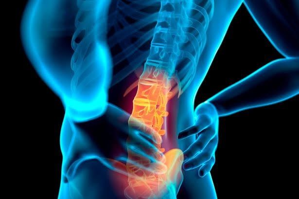
Maybe you have never heard this expression. And is that this disorder is not as well known as osteoarthritis or tendinitis but it is quite frequent, especially in women. Do you feel pain and stiffness? Attentive, because maybe it’s your case.

Physiotherapist in Dwarka calculates that between 3 and 5% of the population will suffer the so-called “frozen shoulder” at some point in their life. Although initially, this percentage may seem low, it increases considerably among certain groups.

80-90% of cases occur in women between 40 and 70 years, especially after menopause (from 50). But they are not the only ones affected. We tell you what this pathology consists of, how to identify its symptoms, which people are more at risk of suffering it, and what treatments exist to overcome it.

WHAT IS THE FROZEN SHOULDER”?


The medical term is adhesive or retractable capsulitis, although the same specialists use the expression “frozen shoulder” because it is much more graphic and easy to understand.

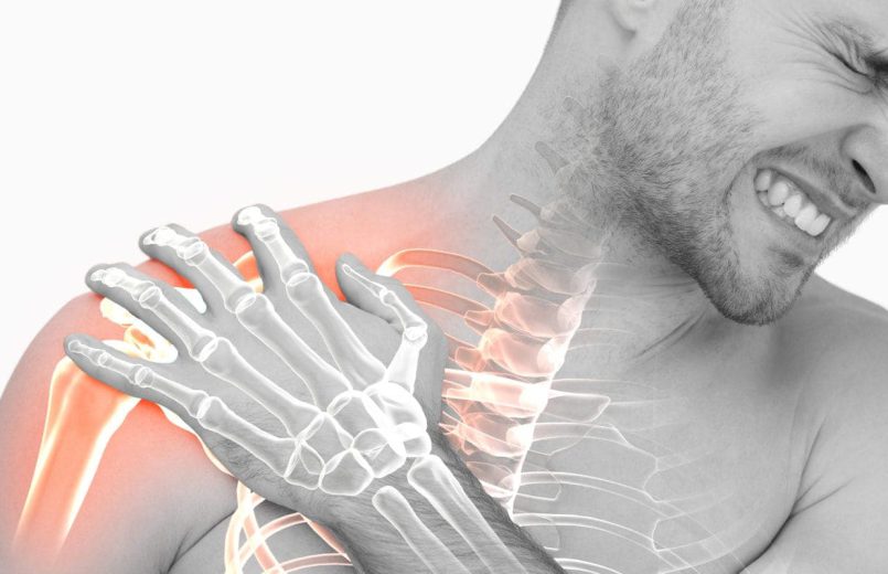
This disorder begins causing a very intense pain in the shoulder that usually gets worse at night. This usually starts suddenly and without a cause that justifies it (bad gesture, injury, etc.).

After a while, the person who suffers begin to notice a stiffness in the joint that ends up in the difficulty or inability to move the shoulder normally. Hence, the term “frozen shoulder.”

The immobility increases progressively until the person is unable to perform such habitual gestures as raising the arm and passing the hand over the head or carrying it towards the back too, for example, fastening the bra, knotting the apron or scratching.

This disorder occurs when there is inflammation in the capsule of the joint, which hinders the mobility of the ligaments that unite the bones that make up the shoulder. However, the cause that causes this inflammation is still unknown.

WHEN THERE IS MORE RISK OF WHICH HAPPENS

Although the reason is not known either, it is demonstrated that certain people are more at risk of suffering from “frozen shoulder”. And is that this disorder is associated with the following factors or pathologies:

  • Hormonal changes. The majority of cases occur in women over 40 years, and especially after menopause.
  • Diabetes. People who suffer from this disease also have a higher risk of suffering from “frozen shoulder”.
  • Thyroid problems Whether you suffer from hypothyroidism or hyperthyroidism.
  • Heart surgery. People who have cardiovascular problems and have undergone open-heart surgery are also among the risk groups.
  • Shoulder injuries Having fractures, surgery on this joint and its immobilization for a while may favor adhesive capsulitis.

If you are in one of these risk groups, you should be alert to the first symptoms, since early diagnosis can help to make the recovery more favorable.

In addition, people with diabetes or with thyroid problems can reduce the risk of suffering “frozen shoulder” if they maintain good control of their disease.

THE TREATMENT DEPENDS ON THE MOMENT

Although in most cases recovery of shoulder mobility is total, the process is very slow and frozen shoulder treatment in Dwarka can last between 6 months and 2 years.

Although sometimes the “frozen shoulder” heals itself after 18-24 months, without treatment and spontaneously, it is advisable to go to the best physiotherapist in Dwarka as soon as possible since the treatment can advance the process, alleviate the symptoms and prevent mobility is reduced.

The physiotherapy doctor in Dwarka will assess the most appropriate treatment according to the phase of the disorder in which the patient is:

  1. Inflammatory phase. The pain is very intense, especially at night, which makes it difficult to rest. But there are still no mobility problems. At this stage, the best physiotherapy doctor in Dwarka usually prescribes analgesics and anti-inflammatories and advises avoiding those movements that may increase pain. Generally, it is advisable to combine a certain rest with gentle stretching exercises. In those acute cases in which the pain does not decrease, it may be necessary to perform some infiltration of steroids (corticosteroids) in the joint.
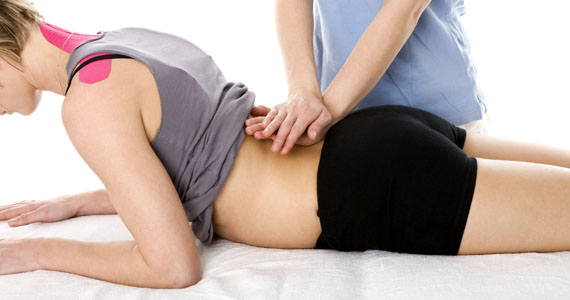
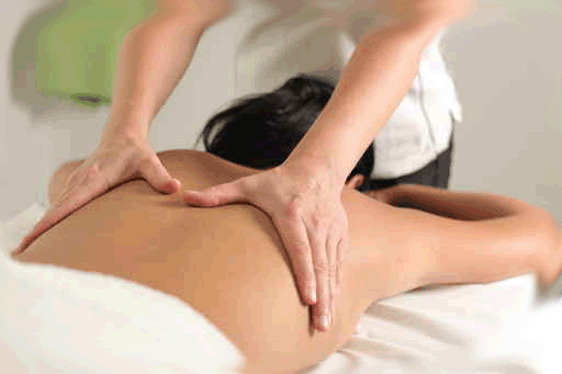
  2. Freezing phase. The shoulder begins to show rigidity and mobility problems are increasing, although the pain usually decreases. At this stage, physiotherapy in Dwarka is fundamental, although perseverance is needed. The number of rehabilitation sessions per week will depend on each case, but they can be daily and for several months. Recovery exercises should always be performed by a specialist physiotherapist in Janakpuri, who will also teach the affected person how to perform the stretching exercises correctly at home.
  3. Recovery or defrosting phase. Mobility progressively improves, although it may be necessary to continue doing exercises for a total recovery and to prevent future relapse.

Most people suffering from “frozen shoulder” respond well to this treatment, as long as they are constant both when going to physiotherapy sessions and when performing the exercises indicated at home.

However, in those cases in which a clear improvement is not achieved, it may be necessary to perform surgery. Normally the operation is performed by arthroscopy in Delhi, through small incisions, and with local anesthesia, and the objective is to “free” the ligaments to restore mobility to the shoulder joint.

DISCARD OTHER PROBLEMS

The shoulder is one of the joints with greater mobility and, therefore, one of the most prone to pain. Therefore, when it does not return in several days it is necessary to go to the orthopaedic in Delhi to make a diagnosis.

In addition to adhesive or retractable capsulitis (“frozen shoulder”), these are the most common problems that can cause pain in this part of the body:

  • Contractures The muscles of the shoulder or cervical muscles contract due to a bad gesture, a bad posture repeated or sustained in time or tension.
  • Calcifications They consist of the accumulation of calcium in the supraspinatus tendon, usually due to minor trauma, tears or inflammation. It usually affects people between 30 and 50 years old.
  • Arthrosis This degenerative disease, in which a loss of cartilage occurs, can also affect the shoulder, as well as other joints such as the knee. In addition to pain and limited mobility, crunches (crepitations) may be felt when moving.
  • Rotator cuff injuries. It is formed by muscles and tendons that provide stability to the bones of the joint and facilitate movement. The tearing or breaking of these tissues by some trauma, sudden movement or wear are some of the most common problems, as well as the inflammation of some tendon (tendonitis).

The symptom of something more serious. When the pain does not vary with the movement, it can be a symptom of some disease that has nothing to do with the shoulder, such as a heart problem (in this case, it is usually accompanied by a feeling of tightness in the chest) or a pulmonary pathology, for example of the pleura. Some types of lung cancer can cause shoulder pain, especially when the tumor is large and press on the area near the joint.