
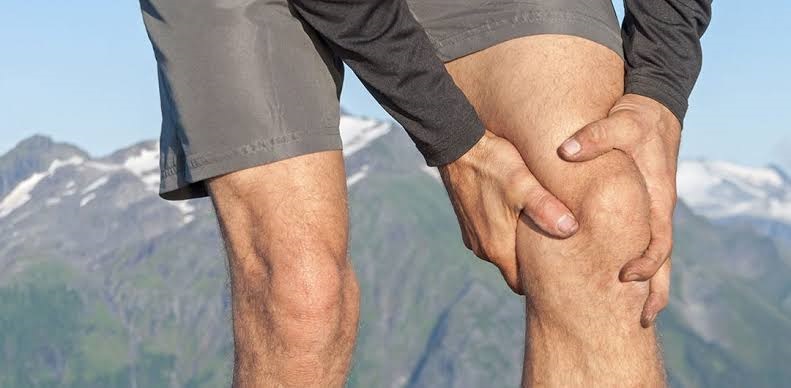
The patellar luxation is a disease of the locomotor system which generally occurs toward the lateral side of the knee, resulting in breakage of the medial patellofemoral ligament (LPFM) in approximately 90-100% of cases.
Although several risk factors have been identified that cause patellofemoral instability after patellar dislocation, appropriate therapy remains a controversial issue.
In this article, we will only talk about the conservative treatment of this lesion.
What is kneecap dislocation?
Label dislocation is defined as the complete loss of contact between the articular surfaces of the patellofemoral joint and in which to restore the normality of the joint a dislocation reduction by a professional with the appropriate knowledge is required, although in In certain cases this reduction occurs spontaneously through a complete extension of the knee.
Types of patellar dislocation: We found three types of patellar dislocation;
- Congenital laxation, which occurs during birth.
- Recurrent dislocation, which tends to recur. A dislocation is considered recurrent when it occurs two or more times.
- Traumatic dislocation, in which there is a loss of contact between articular surfaces in an acute or traumatic way as a result of trauma or forced movement.
The elements involved in the extensor apparatus of the knee are the patella, the femoral trochlea, the quadriceps muscle, the patellar tendon, and the patellofemoral retinaculum.
In the medial retinaculum is the most important ligament in this lesion, the medial patellofemoral ligament (LPFM). This ligament consists of a transverse and an oblique portion and both portions are fused with the vast middle of the quadriceps to be inserted into the patella. This ligament is the main passive stabilizer before the dislocation of the patella during flexion from 0 to 30º.
- The quadriceps angle (angle Q) is a measure of alignment between the femur and the tibia that is measured by drawing an imaginary line that connects the center of the label with the ESIA and a second line that is drawn following the direction of the patellar tendon, from the center of the label to the anterior tibial tuberosity Where these lines intersect, angle Q is formed.
This angle is important since its increase affects the patellofemoral stability and the path of the patella in the trochlea during knee flexion and extension.
- The main risk factors are anatomical, and they are patellar dysplasia, the syndrome of the high patella and the increased distance between the anterior tuberosity of the tibia and the intertrochlear groove (TAT-SIT distance). However, there are other anatomical features that favor patellar dislocation such as ligament hyperlaxity, increased Q angle or atrophy or imbalance of the vast middle of the quadriceps with respect to the vast lateral and a shortened iliotibial waistband.
- The mechanism of injury can be direct, when it is caused by trauma to the medial part of the patella or indirectly when it is caused by a knee flexion movement along with with a knee valgus and internal rotation adduction of the femur, while the tibia abducts and rotates externally.
The indirect mechanism represents 93% of the cases of the dislocated patella.
- For the evaluation and diagnosis of the lesion, in addition to imaging tests such as nuclear magnetic resonance imaging (MRI), radiography or computed tomography (CT), a physical examination must be performed that includes functional tests of the knee (meniscal, varus-valgus, anteroposterior stability, range of motion, etc.) and emphasizing the patellofemoral joint. For this, we will analyze if the sign of the J occurs, which occurs when during the full knee extension, the label moves excessively to the side. In addition, we will perform the patellar apprehension test; With the knee in full extension, we apply a force on the patella directed towards the side and ask the patient to try to flex the knee. If the patient feels pain or dislocation sensation, the test will be positive, indicating femoropatellar instability.
In addition, we will perform a gait analysis to check for possible wrong patterns.
- For the physiotherapeutic treatment of the dislocation of the patella, physiotherapist in Dwarka will begin with immobilization by means of orthotic devices that allow the mobility of the knee, allowing some exercises and a progressive increase in mobility over time. Controlled mobilization reverses the harmful effects of immobilization by stimulating the synthesis and correct alignment of healing tissues so it is important to recover the joint range as soon as possible.
- Treatment of inflammation with cryotherapy, neuromuscular bandage, compression bandage, electrotherapy, soft tissue massage (manual lymphatic drainage), active movement, drugs, and limb elevation.
- Functional or neuromuscular bandage to reduce excessive contact forces in the patellofemoral joint or possible hypermobility of the patella.
- In the early stages of treatment, we can use muscle electrostimulation in addition to an active contraction by the patient to balance the function of the vast middle and the vast lateral quadriceps.
- We must work on the flexibility of soft tissues, especially those that are located in the lateral compartment that, due to their stiffness, can cause instability in the knee; The flexibility of the tensor fascia lata and the iliotibial belt, the vast quadriceps, and the knee flexors should be worked on.
- Balance training and coordination, that is, proprioception exercises, is very important. These exercises cause changes in the nervous system through tasks that seek awareness, sensitization, and enhancement of joint, muscle, tendon and bone structures.
- The most important part of the physiotherapy in Dwarka in the dislocation of the patella is the rehabilitation plan through physical exercise. Once there is a good balance in the extensor mechanism of the knee we will begin a functional training with a gradual increase of the load exercises in the knee.
We will divide the treatment bread into 4 phases:
- First phase: Reduce pain, swelling, and inflammation.
- The second phase: Improve the balance between the vast quadriceps, improve the flexibility of the muscles, re-educate the gait and decrease the excess load in the patellofemoral joint.
- Third phase: Strengthening of the femoral quadriceps and hip muscles and improving coordination and balance in the lower limb achieving a good knee function.
- Fourth phase: Recover the physical and sports level prior to the injury.